PATIENT
PHYSICIANS
Chronic lower limb ischemia plays an important role in the increasing burden of cardiovascular disease. The prevalence of the disease among the population older than 50 years old is 5-8%, and in the presence of risk factors such as hyperlipidemia, smoking, hypertension or diabetes increases up to 30%. Intermittent claudication (IC) affects about 5% of the elderly in the Russian Federation, ie about 2 million people. In 50% of patients the disease may be asymptomatic, 40% suffer from intermittent claudication, 5-10% develop critical limb ischemia (CLI). Thus, the number of patients who need specialized treatment and constant monitoring in the Russian Federation is about 1 million people [AV Pokrovsky, 2013].
Despite the rapid development of vascular surgery in the last three decades, the introduction of new technologies in the treatment of patients with atherosclerotic peripheral arterial disease such as endovascular angioplasty and stenting, the total number of vascular procedures performed in our country is low. According to A.V. Pokrovsky and V.N. Gontarenko, the number of arterial reconstructions in the femoral-tibial area, carried out in specialized departments of vascular surgery, was about 14 thousand in 2012. L.A. Bokeria and B.G. Alekyan’s report on the status of cardiovascular surgery in the Russian Federation in 2012 mentioned 3745 endovascular procedures for the same arterial segment [LA Bokeria, 2012]. Thus, within one year in the Russian Federation less than 18 thousands of endovascular procedures were carried out on atherosclerotic arterial lesions in the infrainguinal segment. According to A.V.Pokrovsky and V.N.Gontarenko, it is necessary to increase the number of surgeries on the arteries by more than ten times. For example, 162 reconstructive procedures on the lower extremity arteries per 100 000 people were performed in the United States in 2007. Only 37 arterial reconstructions per 100 000 population were performed in the Russian Federation in 2012. Thus, we can conclude that the majority of patients with chronic occlusive arterial diseases of the lower extremities, or Peripheral Arterial Disease (PAD), including those with critical limb ischemia (CLI) in the Russian Federation were not able to receive medical aid in specialized hospitals and were treated in general surgical departments [A.V. Pokrovsky, 2013].
One of the new approaches that may be used in the treatment of this group of patients is gene-therapy which is aimed at the induction of growth and development of new blood vessels — angiogenesis. The TASC II (2007) guidelines paid special attention to the provisions relating to gene therapy in PAD. The international committee (Inter-Society Consensus for the Management of Peripheral Arterial Disease) considers that such methods belong to the category of promising treatments efficacy of which has not yet being proven in the CLI treatment, while for the stage II PAD there is some evidence. Russian cardiovascular surgeons and angiologists in the national guidelines for the management of patients with lower extremity arterial disease (2013) determine the benefits of gene therapy and list it as a class IIA evidence treatment for patients with chronic lower limb ischemia. At present, the State register of medicinal products of Russia included the gene therapy drug «Neovasculgen» which contains the supercoiled plasmid containing the gene of Vascular Endothelial Growth Factor (VEGF) as its active component in the list of PAD treatments. Its safety and effectiveness have been studied in multicenter randomized controlled studies, in which the use of “Neovasculgen” lead to a significant improvement in the functional status of patients due to a statistically significant increase in pain-free walking distance, tissue oxygen tension, blood flow velocity, and ankle-brachial index. The drug is intended for combination drug therapy in patients with IIa-III stage of atherosclerotic chronic lower limb ischemia (Pokrovsky-Fontaine). The drug is administered 1.2 mg intramuscularly locally twice at an interval of 14 days.
Registration clinical trial (phases 1-2A-2b and phase 3) was carried out simultaneously in 3 medical centers based on clinical trial approvals (№ 250 dated 03.07.2009 and №177 from 21.04.2010) issued by the Federal Service for the Surveillance in Healthcare and Social Development of the Ministry of Health and Social Development of the Russian Federation; the research protocols were approved by the National Ethics Committee (from 27.05.2009 № 36 and № 62 from 07.04.2010) and the Local Ethics Committee of «Yaroslavl Regional Hospital.» Overall study design, prospective, simple and open was the following: the study included 100 patients (75 in the treated group and 25 in the control group). The drug was administered according to the instructions: local intramuscular injections, as close as possible to the area of ischemia, at doses of 1.2 mg, twice with the interval of 14 days.
The primary efficacy criterion was the distance of Pain Free Walking Distance (PFWD), secondary criteria included ankle-brachial index (ABI), transcutaneous oxygen tension (tcp02), linear blood flow velocity (LBFV). Digital substraction angiography was performed at baseline at chosen time points. The integral results of the treatment were calculated according to the scale of «success» / «failure». The quality of life was determined with the SF-36 questionnaire.
It was found that the PFWD increased by 110%, which was significantly different from the PFWD in control group (P = 0.000). Secondary criteria parameters increased among the patients in the clinical group: ABI value had a 11.11% increase (P = 0.000); TKNK — an 11.38% increase (P = 0.000); LBFV – a 55.12% increase (P = 0.000) compared to control. Angiography performed in patients who received Neovasculgen revealed enhancement of microvasculature contrasting in the affected area due to newly formed collateral vessels as compared to baseline. The patients in the clinical group demonstrated a statistically significant improvement in physical health component (P = 0.000) and tended to have an improved mental health component (P = 0.241). Integrated assessment of the treatment results showed that the frequency of successful outcomes for the period of 6 months in treated patients (94.0%) was significantly higher than in the control group (37.5%; P = 0.000). There were no adverse effects and complications related to the treatment. The results show the overall effectiveness and safety of the drug «Neovasculgen».
38 patients were enrolled in the follow up study after the completion of the registered clinical trials for the additional follow-up examinations at 1, 2, and 3 years [RV Deev et al., 2015]. Safety of the drug was determined by recording all adverse events and serious adverse events, as well as examinations with laboratory and instrumental methods. Screening for cancer among the patients was performed at each visit during the follow up study (3 years). Oncology awareness screening included full examination with ultrasound of abdominal cavity and pelvis, chest X-ray, gynecological examination, electrocardiography, the study of blood coagulation, clinical, biochemical blood tests, and urinalysis. Efficacy criteria were left unchanged: the primary efficacy was pain-free walking distance (PFWD), secondary criteria — transcutaneous oxygen tension (TcPO2), linear blood flow velocity (LBFV) of the affected limb, and ankle-brachial index (ABI). Follow up monitoring of the patients was carried out at Ryazan State I.P. Pavlov Medical Univeristy and the Yaroslavl Regional Clinical Hospital.
There were no adverse events associated with Neovasculgen treatment. No tumor development, impaired vision or other pathological conditions that could indirectly indicate complications of angiogenic therapy were found during the 3-year follow up period . The increase in PFWD in the treatment group was observed during the entire period (Fig. 1); at 3 years an average pain-free walkinf distance increased by 290% ( 390 ± 21 m) as compared to baseline (99.5 ± 11 m).There was a gradual decline in PFWD in the control group: the value decrease by 27% (87 ± 13 m) as compared to baseline. Thus, the difference between the groups in regard to the primary efficacy criterion has reached a statistical significance at 3 months after the start of «Neovasculgen» treatment, whilst PFWD value continued to increase with the maximum recorded at 3 years (p <0,05).
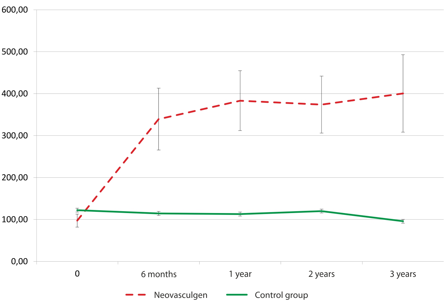
Fig. 1. PFWD dynamics during the clinical trial and 3-year follow-up study
* -differences between treated and control groups were statistically significant (p <0,05)
The dynamics of PFWD increase among the patients treated who received gene therapy was not uniform throughout the observation period. The most signigicat therapeutic effect of the «Neovasculgen» was observed during the first year after application (251% relative to the baseline). During the next 2 years the increase of PFWD was less significant: 49 m on the average (40%). In the control group, by contrast, there were no significant changes in PFWD with its gradual decrease despite the ongoing conservative therapy.
TcPO2 changes were less significant and did not reach statistical significance (fig. 2). The average increase was 13 mm in the treated group by the end of the third year of follow-up study (17%). The positive trend was observed mainly in the 1st year of the observation period; in the following 2 years a stable level of TcPO2 was achieved. In contrast, a gradual decrease in Tcp02 value was detected in the control group ( 3 to 5%).
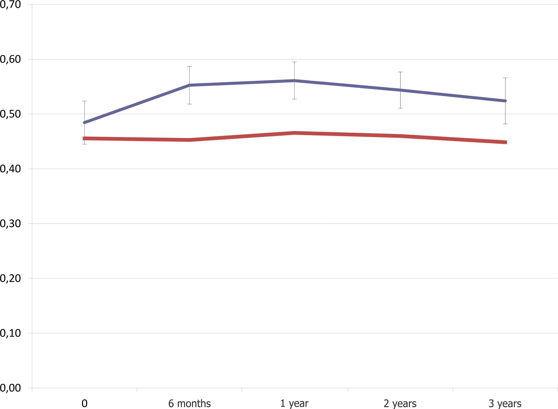
Fig. 2. TcPO2 dynamics during the clinical trial and 3-year follow-up study.
The macrocirculation parameters of affected limb changed slightly throughout the period of observation: there was no statistical significance in regard to ABI level by the end of the 1st year (Fig. 3).

Fig. 3. ABI dynamics during the clinical trial and 3-year follow-up study.
The main goal of the follow up study was to assess the safety of angiogenic therapy with p-VEGF165 gene transfer. There is a traditional awarness of the gene constructions with angiogenic properties due to the role of hypoxia-induced activation of VEGF-VEGFR — signaling system in the genesis of such diseases as cancer or diabetic retinopathy . Thus, there are concerns that the induction of angiogenesis in ischemic tissues of the lower extremities can possibly indirectly stimulate the growth of new blood vessels in tissues distant from the site of gene transfer. However, previous studies showed no signs of increased incidence of cancer, diabetic complications or atherosclerosis progression following the administration of p-VEGF165.
The results of the meta-analysis of multiple trials involving more than 1,000 patients, showed similar incidence of forementioned diseases in the pVEGF and placebo control groups. Moreover, there is data regarding the safety assessments of gene therapy stating that within as long as 8 years after the gene transfer there were no adverse events associated with the induction of angiogenesis. These observations support the results of a 3-year follow-up study of «Neovasculgen» which showed an excellent safety profile, which correlates with the results of global trials in therapeutic angiogenesis. Long-term observation suggests that the pl-VEGF165 gene transfer does not induce the development of malignant tumors or diabetic complications in the long-term period after a standard course of therapy.
The results of the follow-up study have shown that the use of pl-VEGF165 at a dose of 2.4 mg per patient per course resulted in a significant, stable improvement in the functional status of the patients. The therapeutic effect of angiogenic therapy in the clinical group has increased throughout the 3-year observation period, with the most significant increase during the 1st year. On the contrary, the use of standard therapy in the control group was associated with a gradual decrease of the functional parameters in patients, which resulted in 27% PFWD reduction by the end of the 3rd year. Thus, the use of gene therapy in the combination treatment of patients with PAD stage II-III ( Fontaine-Pokrovski) led to a significant clinical improvement when compared to the standard therapy, and prevented the progression of ischemia.
Literature
Deev R., Bozo I., Mzhavanadze N., Voronov D., Gavrilenko A., Chervyakov Y., Staroverov I., Kalinin R., Shvalb P., Isaev A. pCMV-vegf165Intramuscular Gene Transfer is an Effective Method of Treatment for Patients With Chronic Lower Limb Ischemia. J Cardiovasc Pharmacol Ther. 2015.
One of the promising areas of application of the drug “Neovasculgen” is treatment of patients with the Diabetic Foot Syndrome (DFS). The last 20 years of basic research revealed that a major factor in the pathogenesis of ischemia in the DFS is a poor vessel formation, the basis of the granulation tissue growth. Low levels of VEGF in patients with DFS and the activation of BNIP3 (pro-apoptotic protein) enhance catabolic processes along with inhibition of angiogenesis and progression of endothelial dysfunction (Fraisl P., 2009).
According to the results of experimental studies poor vasculogenesis in diabetes can be explained by a decreased progenitor endothelial cells (PEC) mobilization (Kang L., 2009). Several clinical studies showed that there were significantly fewer PEC in diabetic PAD patients as compared to PAD patients who did not have diabetes (Spinetti G., 2005). Poor arteriogenesis, a process aimed at increasing the diameter of collateral vessels in presence of the occlusion of the main vessels was noticed in diabetic patients, which lead to the rapid progression of critical limb ischemia (Walternberger J., 2001). Thus, diabetes impairs multi-component vascular regenerative mechanisms that regulate normal growth and development of blood vessels. In this regard, one of the promising areas of treatment disease in which the formation of new blood vessels plays a critical role, the use of angiogenic therapy is justified, gene therapy in particular. The mechanism of action of gene therapy is based on the transfer and delivery of genes encoding growth factors with angiogenic activity into the cell nucleus to achieve their expression and growth factors production (Losordo D., 2015).
It is known that an increased synthesis of endogenous angiogenic factors in an amount necessary to induce growth and formation of new vessels of the microvasculature, allows to improve perfusion of ischemic tissues. Thus, the use of growth factors is justified in patients with DFS with regard to fundamental mechanisms of vascular growth and development in ischemic conditions caused by the occlusion of major arteries in presence of diabetes. The use of angiogenic therapy in these patients may not only ensure the effective healing of ulcers, but also improve regional perfusion, which may improve limb salvage rates.
The preliminary results obtained from the pilot studies conducted in Ryazan State Medical University, City Hospital 14 of St. Petersburg (Ultimate Rescue Centre), Vidnevskoy Hospital (Moscow Region) and the Regional Clinical Hospital №1 (Khabarovsk) showed the efficacy and safety of the drug «Neovasculgen». The use of “Neovasculge” in patients with DFS led to a significant shortening of the wound healing time, prevented the progression of ischemia and reduces the risk of amputation.
Coronary heart disease (CHD) and its complications continue to be the leading cause of death, despite significant progress in the control of risk factors and treatment, including the increased number of surgical and endovascular revascularization procedures. Surgical treatments showed the advantage compared to the isolated drug therapy which has been demonstrated in large international, multi-center studies. Unfortunately, there is a large group of patients who, for a number of reasons (distal coronary lesions, high pre-operative risk, technical difficulties) can not be a subject of revascularization surgery. Thus, there is a need to find new treatment options for that group of patients with coronary heart disease, who had a poor response to standard medical therapy in order to improve their quality of life, functional status, reduce the risks of complications. Gene therapy uses two basic approaches based on viral and non-viral vectors in order to perform gene transfer and pass a gene construction through the cell membrane into the nucleus. The viral vectors include herpes viruses, adenovirus, adeno-associated virus, lentivirus, and others. Adenoviruses are predominantly used in the treatment of the cardiovascular disease . The results of the first randomized, placebo-controlled study dedicated to the evaluation of the safety and efficacy of gene therapy based on adenoviral vector Angiogenic Gene Therapy Trial (AGENT) were published in 2002 [C.L. Grines, 2002]. The study included 79 participants with a stable angina (functional class 2-3), who were divided into 5 groups, depending on the dosage of angiogenic growth factor (FGF4) delivered via Ad5 vector ( Ad5FGF-4). Monitoring of patients lasted for 311 days after the intracoronary administration. Overall, the results showed safety of the drug, however, a transient rise in body temperature was observed within 24 hours after the administration in 3 patients in the group who received the highest dose, and yet in another 2 patients there was a significant increase in the activity of the liver enzymes in the blood, Another three randomized placebo-controlled studies which evaluated the safety and efficacy of Ad5FGF-4 (AGENT-2, AGENT-3, AGENT-4) were conducted later [T.D. Henry, 2007]. A meta-analysis of these studies did not originally identify any benefits of Ad5FGF-4 transfer, however, the re-analysis of the effectiveness among groups, divided by gender, showed a statistically significant improvement in tolerance to physical activity rates among women.
In another randomized REVASC trial (Randomized Evaluation of VEGF for Angiogenesis) the direct intramyocardial injection of adenoviral construction containing VEGF 121 gene was studied. The drug was administered after a mini-thoracotomy directly into the myocardial wall [D.J. Stewart, 2006]. The study enrolled 67 patients with stable angina (functional class 2-4) in the presence of hemodynamically significant stenosis in at least two coronary arteries. The patients were divided into two groups: conservative therapy, and direct injection of 1010 AdVEGF121.The incidence of serious adverse events during the follow-up period was similar in both groups. The results obtained after 26 weeks following the endocardial injection showed significant improvement in the exercise tolerance in patients who received AdVEGF121. However, the single-photon emission computed tomography showed no significant improvement in the perfusion of ischemic myocardium. Unfortunately, the greatest disadvantage of this study was the absence of a placebo group, which did not allow to conclude whether the improvement of the functional status of patients who received AdVEGF121 was the result of gene-therapy, and not a placebo effect. Another approach to gene therapy is the use of nonviral plasmid vectors. A plasmid is a small circular DNA molecule, which has the ability to penetrate the cell nucleus and replicate autonomously. Due to such properties a plasmid can exert an effective transfer of sequences with therapeutic genes into the nucleus. Plasmids were studied in experimental trials: the results have shown that plasmid vectors had considerably less significant transfection properties relative to viruses, however, the expression of the genes inserted into the plasmid lasted for longer periods of time, thus maintaining the desired concentration of growth factor suitable for formation and maturation of new vessels. The use of plasmid vectors was not accompanied by the development of immune reactions, which considerably suspended the possible use of viral vectors uses.
Gene-therapy drugs based on non-viral vectors are used significantly more often, since they do not possess cytotoxic properties, like adenoviral ones. Experimental and clinical studies have shown certain side effects arising from the use of adenoviral vectors including a transient rise in body temperature after injections, increase in the concentration of hepatic enzymes, and the C-protein level. One of the main advantages of plasmid vectors is their rapid degradation by different enzymes in the bloodstream. The use of plasmid vectors helps to prevent the risk of systemic drug effects such as those with viral vectors, which are able to induce the synthesis of a target protein far away from the place of the intended therapeutic effect. Finally, the manufacturing process of potential drugs based on non-viral vectors is significantly less expensive and easier technically.
Thus, the advantage of the use of viral vectors is their high transfection activity, which allows to achieve high level of gene expression of the therapeutic agent. However, several studies have demonstrated that the level of expression of the growth factor can reach its maximum, and at a certain concentration might further increase the transfection efficiency to a level that may be toxic.
The first clinical study designed to assess the efficacy and safety of a plasmid DNA containing VEGF gene was conducted by J. Isner and D. Losordo in 1998 [J. Isner, 1998]. A small pilot study involved 5 patients with stable angina functional class 3-4. For a number of reasons it was impossible to perform revascularization surgery in those patients, and the conservative therapy was ineffective. pl-VEGF at dosage of 125 micrograms was administered after the mini-thoracotomy directly into antero-lateral left ventricular wall. No side effects during the procedure or side effects in the postoperative period were observed. The single-photon emission computed tomography performed 60 days after the administration of pVEGF showed that there was an improvement in perfusion of the ischemic infarction area, which correlated with the improvement of the functional status of the patients, including a decrease in the functional class of stable angina, reduced number of angina episodes during the week, and the reduced need to use nitroglycerin. Thus, the preliminary positive results were obtained from this small study. A phase 1 clinical trial was conducted in order to evaluate the safety and effectiveness of the direct gene transfer with a plasmid DNA , which contains a gene encoding VEGF in patients who were not candidates for revascularization surgery by performing a mini-thoracotomy. The study included 20 patients with coronary heart disease, stable angina functional class 3-4; the subjects were divided into two randomized groups depending on the dosage of pVEGF, 125 µg or 250 µg. The results showed an improvement in the functional status of the patients, which was reflected in a decrease of the angina class. According to the single-photon emission computed tomography, an improvement in ischemic areas of the myocardial perfusion was detected at 60 days after endocardial injection procedure. However, the design of the study did not allow a placebo-control to determine the proper effectiveness of a plasmid DNA encoding VEGF. In general intracoronary administration of the gene therapy construction was inefficient due to its rapid disintegration in the bloodstream. The situation changed with the invention of modern methods of drug delivery, based on electromechanical mapping of the heart. A good example is the electrophysiological navigation NOGA XP system. Due to the presence of a special sensor at the end of the catheter it forms a three-dimensional spatial structure of the left ventricle after being placed into the cavity of the left ventricle, which gives a possibility to visualize ischemic lesions and deliver the drug directly to the zone with reduced perfusion. For the first time the feasibility of this approach was demonstrated in 1999 by J. Isner and D. Losordo [D.W. Losordo, 1999]. The patient was a 82 year old male with coronary heart disease, stable angina class 3, who had previously had a coronary artery bypass grafting without clinical effect. Transendocardial injections of a plasmid DNA containing the VEGF were performed. The sites of injections were chosen according to the electroanatomical mapping of selected areas with reduced unipolar voltage and local linear reduction. In 2001, these same researchers have conducted the first randomized, placebo-controlled pilot study of the safety and efficacy of intracardial injections of a plasmid DNA with the VEGF-2 gene. 200 micrograms of pl-VEGF2 were injected in the areas of hibernating myocardium of six patients with coronary heart disease, stable angina class 3-4, using the navigation system (NOGA XP). An imitation of endomyocardial injection procedure was performed in patients in the placebo group. The study results showed no complications during administration and during the follow-up period, patients’ functional status in clinical group was significantly improved, causing a decrease in the number of angina episodes and nitroglycerin use. According to the single-photon emission tomography performed 90 days after the endomyocardial injection procedures, patients in clinical group had an improvement in the perfusion of ischemic areas of the myocardium, as compared to the placebo group.
Later a few small clinical studies with different design were performed, aimed at assessing the efficacy and safety of endomyocardial administration of a plasmid DNA with VEGF gene using the navigation NOGA XP system. However, the results of these studies did not allow to make an unambiguous conclusion about the effectiveness of this approach.
The first large, placebo-controlled clinical study was EUROINJECT-1, the results of which were published in 2005. The study involved 80 patients with coronary heart disease, stable angina functional class 3-4. Patients were divided into two groups of 40 subjects each. In the treatment group the plasmid DNA with VEGF gene was administered in a dosage of 500 micrograms, the placebo group of patients received a plasmid DNA containing no genetic sequences. The research results were obtained at 3 months and showed no statistically significant difference in the change of the functional status of patients, whether a decrease of functional class of stable angina or an increase in exercise capacity. However, there was a significant improvement in perfusion according to the single-photon emission computed tomography and an improvement in the contractile function of the left ventricle according to the electromechanical mapping and echocardiography.
The next major study — NORTHERN (The NOGA angiogenesis Revascularization Therapy assessment by Radio Nuclide imaging) was conducted in 2009. The study involved 93 people with coronary heart disease, stable angina functional class 3-4, who were not candidates for revascularization surgery. Patients were randomized into two groups: VEGF gene transfer was performed via 10 injections of 0.2 ml into the hibernating myocardium zone at a total dose of 2000 mg; a buffer solution was used in the placebo group. The primary endpoint in this study was an improvement of perfusion according to the single-photon emission computed tomography; the secondary efficacy criteria were indicators of the functional status of the patients . Unfortunately, there were no statistically significant differences with regard to primary criterion despite the relatively high dose of the drug; several secondary efficacy criteria demonstrated the benefit of using a plasmid DNA with VEGF gene as compared to placebo group.
Thus, there are inconsistent results regarding the efficacy of a plasmid DNA containing VEGF gene. Unfortunately, it is impossible to make definitive conclusions on versatile results of different clinical trials, which significantly differed in design. The results varied depending on the availability of the placebo group, which allowed to control some of the psychological aspects associated with participation in a clinical trial. Another component was the mandatory use of «blind» trials, which greatly reduced the likelihood of a deliberate distortion of the results. Discrepancy of the research findings can be explained not only by the different study design, but also by such factors as careful selection of patients and rational choice of performance criteria. The second group of causes related to the structure and design of the plasmid based gene constructions. Previous publications only mention the use of a plasmid DNA containing VEGF gene, and do not indicate its exact codon sequence, promoter, and other important aspects of drug design, which may have a significant impact on the gene transfection and the efficiency of its expression.
We have conducted a preclinical study of the effectiveness of «Neovasculgen» in the experimental model of acute myocardial infarction in order to obtain preliminary information and a basis to conduct a clinical study. A week after the ligation of the left coronary artery the experimental group of mice was treated with pl-VEGF gene transfer in a dose of 200 micrograms. Parameters of the left ventricular function were assessed using MRI and echocardiogram as the main performance criteria. Afterwards, the necessary material was obtained from the animals and studied using histological and immunohistochemical analysis in order to evaluate the effect of angiogenic therapy on necrosis areas and the number of capillaries per unit area. The results showed that administration of the pl-vegf165 improved the functional parameters, which correlated with the increased the number of vessels. Thus, therapeutic angiogenesis via gene transfer had positive results in an experimental model of acute myocardial infarction in animal study, such as better vascularization of the peri-infarction areas which, apparently, helped optimize the recovery of myocardium both morphologically and functionally, which was not a subject to irreversible damage during a heart attack (necrosis). Thus, the results of preclinical studies have allowed us to design a clinical trial to assess the efficacy «Neovasculgen» in patients with coronary heart disease.
More details on the results of pre-clinical studies:
According to various data, the incidence of peripheral nerve damage is 3-10% [A.R. Hudso, 1977]. The necessity to develop treatments for the peripheral nerve damage is hard to overestimate. New treatment options may improve the quality of life, contribute to the standard care for such patients, shorten the length of the recovery period (a year or more at the moment), cause a significant improvement in the quality of life of patients of working age, and decrease disability rates. Peripheral nerve injury is a frequent cause of disability leading to job loss. The peripheral nerve damage is caused by a number of factors: injuries of different mechanism, the time elapsed from the time of injury before the surgery, the length of the peripheral nerve injury, etc. One type of reconstructive treatment is stitching the stumps of a transected nerve by creating an end-to-end anastomosis. However, the injury of the peripheral nerve often leaves the defects of different length, so that anastomotic approach is not always possible. In this situation, the «gold standard» for restoring the integrity of the nerve is to use autologous nerve insert. A functionally less significant nerve can be used as an autograft. Alternatively, a variety of tubular structures may be used to eliminate an extended defect of tissues and create favorable conditions for the regeneration of peripheral nerve. The extent of the recovery of the limb function after surgical repair of peripheral nerve integrity depends on many factors: the time elapsed since the injury prior to the operation, the length of the defect, the distance from the site of peripheral nerve injury to the innervation area, etc. However, despite the development of technologies aimed at restoring the nerve integrity even in hostile environment, in the end there is often limited recovery of the limb function. It encourages the research of new treatments that would improve the standard of reconstructive treatment and quality of life of patients. One of the possible approaches is the use of growth factors to induce peripheral nerve regeneration. This concept was the result of the accumulation of data regarding the significant role of growth factors in the natural process of regeneration of peripheral nerves [G. Terenghi, 1997].
VEGF is one of the most well-studied growth factors which affect the recovery of peripheral nerves. For a long time after its discovery VEGF was considered as an inducer of angiogenesis and a potential therapeutic agent for the treatment of various diseases associated with tissue ischemia. However, some data on its neuroprotective properties were shown for neurons of the peripheral and central nervous system [M. Sondell, 1999]. VEGF induces proliferation of Schwann cells, astrocytes, microglia, neurons of the cerebral cortex [Zhu, Y., 2003]. In rat models of the intersection injury of the sciatic nerve there was a significant increase in VEGF expression and Flt-1 (VEGF II receptor type) expression in the spinal cord as well as in the lumbar spine in response to injury [R.R. Islamov, 2004]. Thus, there were certain premises for the application of the growth factors as a component of the therapeutic reconstructive treatment of peripheral nerve injuries. The use of a VEGF composition with a Matrigel filling induced axonal sprouting, which caused an increased number of axons in the cross-sectional area [J.M. Rovak, 2004]. The use of polylactic acid pellets with VEGF in the grafts in the model of peripheral nerve injury with long defects in peroneal and tibial nerves, significantly improved the function of the nerves and increased the number of myelinated fibers in the grafts [H. Karagoz, 2006].
VEGF induced the division and migration of Schwann cells along the experimental graft, which correlated with the increase in the number of capillaries and myelinated fibers [14]. Introduction of VEGF in combination with another growth factor BDNF into the cavernous body in a rat model of cavernous nerve injury resulted in restoration of the lost function and erectile innervation [P.S. Hsieh, 2003].
However, the use of growth factors for therapeutic purposes has several limitations. When administered in the damaged area, they undergo rapid degradation, as a consequence, there are some difficulties in maintaining a constant concentration of factors needed to achieve the desired therapeutic effect [P. Haninec, 2012]. Accordingly, there is an increasing interest in the use of gene therapy. Transfer of a gene encoding a therapeutic agent has two main options: the use of viral vectors and non-viral plasmids. However, the use of viral vectors in the clinical application is limited (despite the high transfection rates) because of the risk of insertional mutagenesis and potential toxicity of the inflammatory response. A more secure method of the gene transfer is the use of plasmid DNA. In the model of reconstruction of the musculocutaneous nerve using an end-to-end and end-to-side anastomosis, an intraoperative administration of a plasmid DNA comprising a sequence of VEGF gene in the distal stump, resulted in a significant increase in the number of myelinated fibers in the cross-sectional area distal to the anastomotic site, which correlated with a significant increase in the concentration of VEGF in Schwann cells. Similar results can also be achieved with paraneural injection. In the model of sciatic nerve injury pl-VEGF was administered intramuscularly and was used in combination with hyaluronic acid films that covered the anastomosis to reduce the severity of scarring process. Intramuscular administration of the fore mentioned drug was associated with a significant increase in the amplitude of the muscular response and an increase in the number of myelinated fibers in anastomotic area [F. Zor, 2014]. The study conducted by Wang F. et al. a dose-dependent effect of the administration of pl-VEGF was shown after crosslinking the stumps of the sciatic nerve with an end-to-end anastomosis. High doses of pl-VEGF provided for the most significant increase in neurophysiological parameters. Thus, the experimental studies demonstrated the possibility of using gene constructions to improve peripheral nerve regeneration and were prerequisite for clinical studies to assess the possibility of using this approach for the treatment of peripheral nerve injury in humans.
Currently, we conduct a pre-clinical study of the efficacy of the drug «Neovasculgen» in traumatic peripheral nerve injury, accompanied by its complete intersection . The data obtained by this moment shows preliminary evidence suggesting that intraneural injection of the genetic drug provides a statistically significant shortening of the recovery time according to the results of electrophysiological and functional performance tests, which creates the basis for the improvement of regenerative conditions, and for the axonal growth induction. The results of the experimental have allowed us to plan a clinical trial (NCT02352649) regarding the effectiveness of the drug «Neovasculgen» in patients with peripheral nerve injury.


